
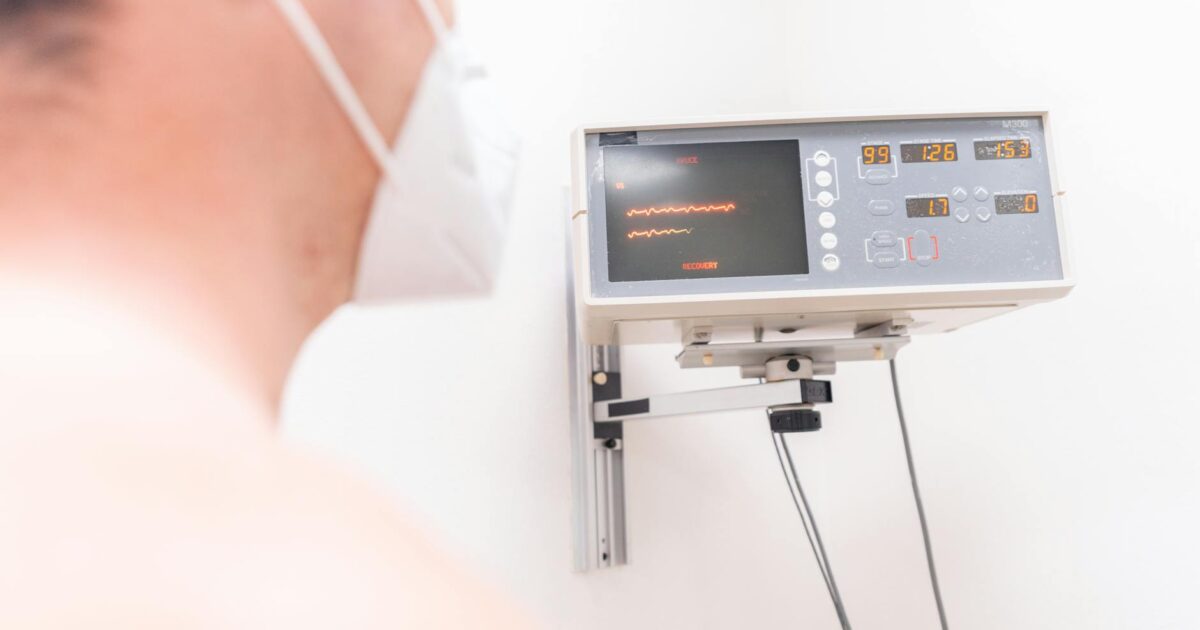
Understanding the intricate interplay between the heart and lungs offers profound insights into a patient’s physiological state, particularly in critical care settings. Among the most valuable of these insights is the measurement and interpretation of pulse pressure variation (PPV), a dynamic parameter that plays a key role in guiding fluid management strategies as of April 2026. This tool provides real-time information about how a patient’s cardiovascular system responds to mechanical ventilation, directly influencing decisions regarding fluid administration.
Last updated: April 26, 2026
What is Pulse Pressure Variation?
At its core, pulse pressure refers to the difference between systolic and diastolic blood pressure. While a single pulse pressure value offers a snapshot, it’s the variation in this pressure over the respiratory cycle that truly unlocks its diagnostic potential. Pulse pressure variation quantifies these breath-by-breath changes, reflecting the heart’s stroke volume responsiveness to alterations in intrathoracic pressure. This dynamic measurement moves beyond static indicators like central venous pressure, which have demonstrated limited predictive value for fluid responsiveness, offering a more accurate assessment of a patient’s fluid needs.
The Physiology Behind PPV
The physiological basis of pulse pressure variation involves the constant interaction between the respiratory and circulatory systems. During mechanical inspiration, positive pressure increases intrathoracic pressure, compressing the vena cava and reducing venous return to the right heart. This decreases right ventricular preload, leading to less blood pumped into the pulmonary circulation. Consequently, left ventricular preload, stroke volume, and pulse pressure are reduced. Conversely, during expiration, intrathoracic pressure falls, allowing increased venous return, which augments stroke volume and pulse pressure. The magnitude of this difference between inspiratory and expiratory pulse pressures, expressed as a percentage, defines PPV. As of 2026, this physiological principle remains fundamental to understanding PPV’s utility.
Clinical Application: Guiding Fluid Responsiveness
Clinicians use pulse pressure variation as a key indicator for determining fluid responsiveness in mechanically ventilated patients. A high PPV, typically considered above 10-13% as of April 2026, suggests the heart is operating on the steep portion of the Frank-Starling curve. This implies that administering fluids is likely to significantly increase cardiac output and stroke volume. Conversely, a low PPV, generally below 10%, indicates the heart is well-filled and operating on the flat portion of the curve. In such instances, additional fluids may not improve cardiac output and could lead to detrimental fluid overload, potentially exacerbating conditions like pulmonary edema or acute respiratory distress syndrome (ARDS).
The dynamic nature of PPV allows for continuous assessment, enabling clinicians to adjust fluid therapy in real-time as a patient’s condition evolves. This is particularly beneficial in complex scenarios where fluid needs can change rapidly, such as during sepsis management or in the post-operative period following major surgery.
Broader Clinical Utility and Recent Developments
The utility of pulse pressure variation extends across various intensive care units and operating rooms in 2026. For example, during major surgery, anesthesiologists utilize PPV to optimize fluid administration, ensuring adequate tissue perfusion while avoiding excessive fluid buildup, which can complicate recovery. In medical ICUs, it aids in guiding resuscitation for patients in septic shock, a critical application that helps prevent both hypovolemia and hypervolemia. Personalized fluid management, informed by real-time data like PPV, contributes to improved patient outcomes, a trend strongly emphasized in critical care guidelines updated in late 2025.
Recent research continues to refine our understanding of PPV. Studies published in 2024 and early 2026 have explored the hemodynamic determinants of pulse pressure itself, offering a deeper understanding of the underlying physiological mechanisms. According to research published in journals such as JAMA Network Open (February 15, 2026), further investigation into the predictive value of PPV for microcirculatory blood flow impairment is ongoing, highlighting its potential beyond just macrohemodynamic assessment. This aligns with ongoing efforts to refine fluid management strategies, particularly in challenging populations like pediatrics, where fluid responsiveness remains an active area of research, as noted by publications in Pediatric Critical Care Medicine (November 2025).
Furthermore, as reported by Anesthesiology News (March 10, 2026), advancements in ventilator technology and integrated monitoring software now allow for more sophisticated analysis of PPV, even in the presence of some arrhythmias or less ideal ventilation patterns. These systems can often provide more nuanced interpretations, prompting further clinician review rather than simply presenting a single numerical value.
Limitations and Considerations for Accurate Use
Despite its significant advantages, it’s essential for healthcare professionals to understand the limitations of pulse pressure variation in 2026. Its accuracy depends on specific patient and ventilator conditions: the patient must be mechanically ventilated with controlled ventilation, and critically, free from spontaneous breathing efforts. The tidal volume must be sufficiently large, typically recommended at 8 mL/kg of ideal body weight or more, to induce meaningful intrathoracic pressure changes that PPV relies upon. Failure to meet these criteria can lead to falsely low or absent PPV values, even in fluid-responsive patients.
Several clinical conditions can also affect the reliability of PPV. These include cardiac arrhythmias (like atrial fibrillation), open chest procedures, significant right ventricular dysfunction (which can decouple preload and afterload), severe hypovolemia or hypervolemia, increased intra-abdominal pressure, and very low lung compliance (as seen in ARDS or severe pneumonia). In these scenarios, PPV may not accurately reflect fluid responsiveness, and clinicians must integrate this data with other hemodynamic parameters and clinical assessments. As highlighted in a recent review in Critical Care Medicine (January 2026), pulse pressure amplification, a related but distinct parameter reflecting arterial waveform changes, has also been identified as a hemodynamic predictor of cardiovascular disease, underscoring the complexity of vascular dynamics that must be considered alongside PPV.
Technological Integration and Future Directions
Modern physiological monitoring systems, including advanced ventilators and bedside monitors, increasingly incorporate automated calculations of pulse pressure variation. This technological integration makes PPV readily available at the bedside, reducing manual calculation errors and enabling more timely, evidence-based decisions. This shift moves away from subjective assessments towards precise, data-driven fluid management.
The continued integration of dynamic parameters like PPV into routine clinical practice signifies a broader shift towards more precise and personalized medicine in critical care as of 2026. Ongoing research is exploring its predictive capabilities further, such as its potential role in predicting outcomes in patients with specific types of shock or its utility in non-invasively monitored patients using advanced algorithms. The evolution of artificial intelligence (AI) in healthcare is also expected to enhance the interpretation of PPV, potentially identifying subtle patterns and integrating it with a wider array of physiological data for more comprehensive hemodynamic assessment.
Latest Update (April 2026)
As of April 2026, the focus in PPV research and application is increasingly on its integration with other dynamic indices and advanced monitoring technologies. New algorithms are being developed to improve PPV’s accuracy in patients with less predictable respiratory mechanics, such as those undergoing lung-protective ventilation strategies with lower tidal volumes. Furthermore, studies are investigating the correlation between PPV and tissue oxygenation markers, aiming to bridge the gap between macrohemodynamic status and microcirculatory perfusion. The consensus among critical care experts, as reflected in recent society guidelines from late 2025, is that while PPV is a powerful tool, it must always be used within a comprehensive clinical context, considering the patient’s overall condition and other available data.
Frequently Asked Questions
What is the primary benefit of using PPV in patient management?
The primary benefit of using pulse pressure variation (PPV) is its ability to dynamically predict fluid responsiveness in mechanically ventilated patients. It helps clinicians decide whether administering intravenous fluids is likely to improve cardiac output, thereby optimizing tissue perfusion and potentially improving patient outcomes, while also helping to avoid unnecessary fluid administration and its associated risks.
Under what specific conditions is PPV most reliable?
PPV is most reliable when the patient is fully mechanically ventilated with a consistent tidal volume (at least 8 mL/kg ideal body weight), has no spontaneous respiratory efforts, and is in a regular sinus rhythm. Conditions like arrhythmias, spontaneous breathing, or open chest surgery can significantly reduce its reliability.
Can PPV be used in spontaneously breathing patients?
Traditionally, PPV requires controlled mechanical ventilation. However, modified techniques and related parameters like stroke volume variation (SVV) calculated from arterial lines or pulse contour analysis are sometimes used or adapted for spontaneously breathing patients, though their reliability may differ and require careful interpretation and validation with other clinical signs.
What is considered a significant PPV value indicating fluid responsiveness?
As of April 2026, a PPV value generally above 10-13% is considered indicative of fluid responsiveness in appropriate clinical settings. However, the exact threshold can vary slightly depending on the specific patient population, the monitoring equipment used, and the clinical context. Clinicians often look for trends and interpret the value alongside other physiological data.
Are there any newer technologies or methods that improve upon traditional PPV?
Yes, ongoing advancements include integrated algorithms in modern ventilators that can automatically calculate PPV and SVV, potentially offering more consistent measurements. Research is also exploring the use of advanced signal processing and AI to enhance PPV’s accuracy in more challenging patient scenarios, such as those with suboptimal ventilation or arrhythmias, and to correlate it more directly with microcirculatory parameters.
Conclusion
Pulse pressure variation remains a valuable dynamic predictor of fluid responsiveness in mechanically ventilated patients as of April 2026. Its ability to provide real-time, beat-to-beat data on the heart’s response to ventilation-induced changes in preload allows for more precise fluid management. While it has limitations and requires careful application within specific clinical conditions, understanding and correctly applying PPV significantly enhances a clinician’s ability to optimize hemodynamic status, improve oxygen delivery, and ultimately contribute to better patient outcomes in critical care settings.
Sabrina
2 writes for OrevateAi with a focus on agriculture, ai ethics, ai news, ai tools, apparel & fashion. Articles are reviewed before publication for accuracy.


